
LESSON 1
THE MAMMALIAN EYE
The mammalian eye is a receptor organ responsible for sight. It contains photoreceptor cells, which perceive the light stimulus and change it into nervous impulse.
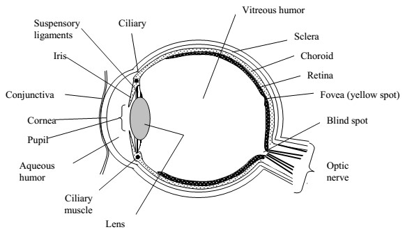
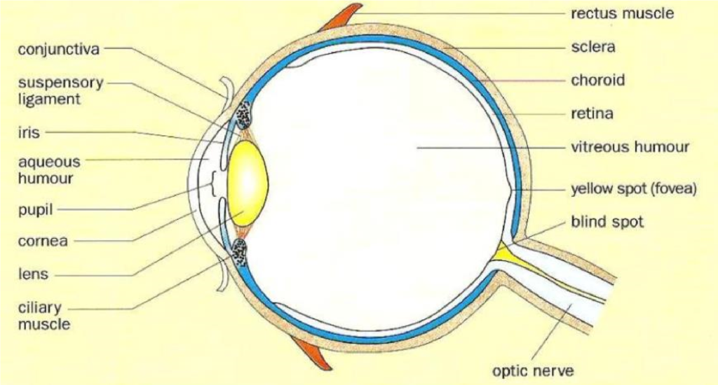
Parts of the eye
1. The conjunctiva:
This is a thin transparent layer lining the inside of the eyelid.
It protects the eye and holds it in position.
It enables the eye ball to move easily by secreting mucus.
2. The sclera:
This is a tough inelastic layer that gives shape to the eye.
It protects the inner most delicate parts.
It provides attachment for the muscles of the eye.
3. The cornea:
This is a transparent layer in front of the eye.
It refracts (bends) light into the eye.
4. The choroid layer:
It is below the sclerotic layer.
It is pigmented and mainly contains black pigment which stops reflection of light rays.
It prevents internal reflection of light.
This contains a network of blood vessels supplying oxygen and food to the eye.
5. The aqueous humour:
It is a solution of sugar, salts and proteins.
The aqueous humor is a watery fluid which maintains the shape of the eye. It also refracts light into the pupil and the lens.
6. The vitreous humuor:
It is a jelly-like substance that fills the inner cavity of the eye.
It is transparent and maintains the shape of the eye. It refracts light to the retina.
7. The ciliary body:
This contains ciliary muscles, which control the size of the lens during viewing nearby or distant objects.
8. The lens.
It is transparent and held by suspensory ligaments.
It refracts light to make an image on the retina.
9. The iris
This is made up of an opaque tissue the center of which is a hole called pupil that allows in light to form an image on the retina.
The contraction of the muscles of the iris increases the size of the pupil and relaxation decreases the size of the pupil.
It is therefore responsible for controlling the amount of light entering the eye.
10. The retina
This is a layer containing photoreceptor cells (light sensitive cells)
There are two types of light sensitive cells on the retina
i) Rods
ii) Cones
The cones are sensitive to coloured light and are responsible for colour vision. They are also sensitive to light of high intensity and are used during daytime.
Most cones on the retina are concentrated on the fovea or yellow spot. The rods are incapable of perceiving coloured light and are sensitive to light of low intensity (dim light). They are used during night vision.
Nerve fibers from the photoreceptor cells run to the brain via the optic nerve. The rods contain a pigment rhodopsin which is rapidly bleached by even a small amount of light but at the same time it is rapidly generated.
The cones contain a pigment called iodopsin which is less sensitive to light and is not bleached so quickly.
The retinas of nocturnal animals have mainly rods. Due to this, nocturnals can’t perceive different colours.
Therefore the retina is where the image is formed in the eye.
11. Pupil
This is a round black hole in the center of the eye lying behind the cornea. It allows light to pass into the eye to the lens.
12. Suspensory ligaments.
These are inelastic fibers that hold the lens in position.
13. The blind spot:
This is a region where the nerve fibers leave the eye to enter the optic nerve. It has no light sensitive cells. When an image falls on this point, it is not taken to the brain thus blind spot.
14. The fovea
This is a small depression in the center of the retina. It has only cones in a high concentration. It is therefore a region on the retina that contains the largest number of sensory cells. Due to this, it produces the most accurate images in the eye.
15. Eye lids
These protect the eye and remove any foreign bodies that enter it. Regular blinking enables the spread of the fluid all over the exposed surface of the eye.
16. Eye lashes
They prevent dust particles and other objects from entering the eye.