
LESSON 5
THE EAR
Structure of the ear
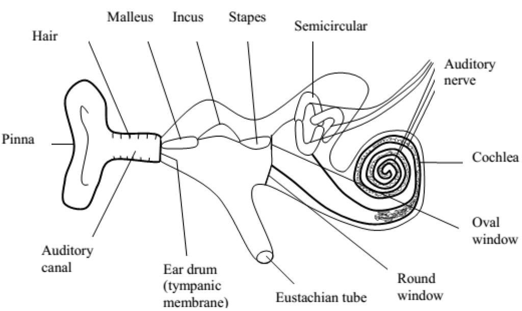
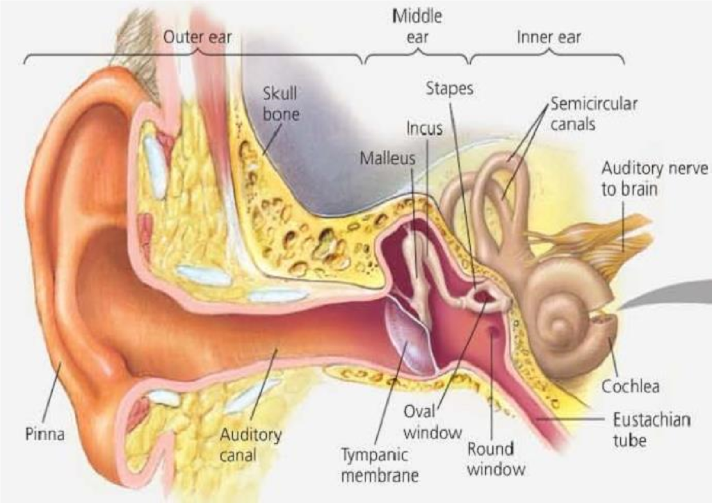
The ear has sensory receptors for hearing and balancing. These are mechanoreceptors because they respond to pressure and gravity. The ear is made up of three areas i.e. the outer ear, middle ear and inner ear.
1. The outer ear:
This is the tube opening to the side of the head and inwards stopping at the eardrum. It has an outer extension called the pinna. The pinna concentrates and directs the sound vibrations into the ear through the auditory canal. This makes the ear drum to vibrate.
2. The middle ear:
This is a cavity in the skull filled with air. It communicates with the mouth cavity through the Eustachian tube. There are three small bones called ossicles in the middle ear which link the eardrum and the opening of the skull called oval window that leads to the inner ear.
3. The inner ear:
The inner ear is filled with a fluid and consists of mainly a coiled tube known as the cochlea. The cochlea has sensory nerve endings leading to the brain. These transmit nervous impulses from the ear to the brain.
Functions of parts of the ear
1. Pinna:
2. Ear ossicles:
These are 3 tiny bones in the middle ear. They are:
- Malleus (hammer)
- Incus (anvil)
- Stapes (stirrup)
They are joined like a chain and they transmit sound vibrations across the middle ear from the ear drum to the oval window. They amplify sounds of low tones.
3. Eustachian tube:
- It connects the middle ear to the pharynx of the mouth.
- Its function is to equalize air pressure on both sides of the ear drum so that it can vibrate freely.
- It opens when one is swallowing and yawning.
- It prevents the eardrum from bulging.
- The Eustachian tube is used to balance the pressure inside the ear with that outside the ear.
- When the pressure of air in the middle ear is higher than that of the atmosphere, yawning takes place and air escapes from the middle ear through the Eustachian tube to the mouth where it is lost. This reduces the pressure back to normal.
- When the pressure of air in the middle ear is lower than that of the atmosphere, yawning takes place to allow the atmospheric air to go into the middle ear through the mouth and Eustachian tube. This raises the pressure back to normal.
4. Oval window (fenestra ovalis):
It is a flexible membrane which vibrates and sets up vibrations in the fluids of the ear called perilymph in the cochlea.
It receives impulses from the steps and transmits them to the cochlea.
5. Round window (fenestra rotunda):
It is a flexible membrane which controls the displacement in the cochlea created by vibrations of ossicles by releasing pressure when it bulges out wards.
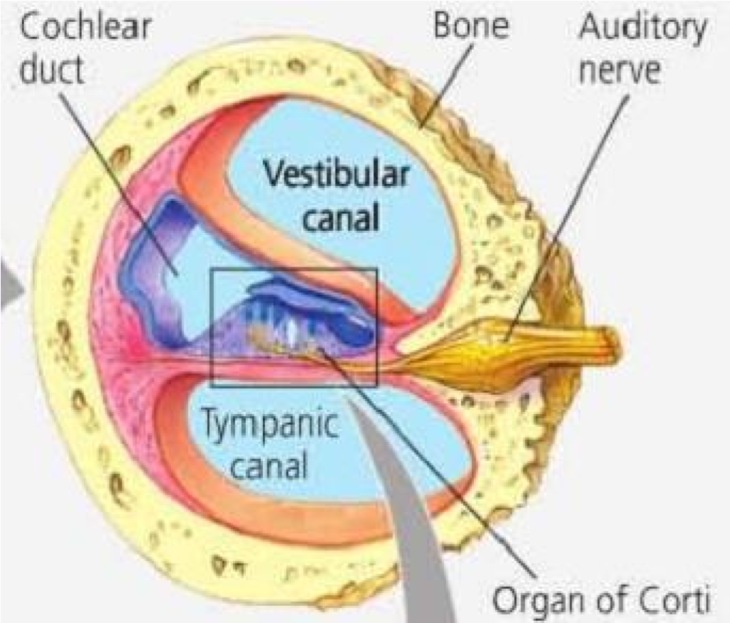
6. Cochlea:
It is a 3 chambered fluid filled tube which is coiled. It contains sensory cells which pick up vibrations in the fluid and transmit them to the auditory nerve. The sound vibrations move along the auditory nerve and reach the brain where they are interpreted as sound. Its 3 chambers include:
(i) Vestibular canal (scala vestibuli)
It is the upper canal which starts from the oval window. It contains perilymph.
(ii) Tympani canal (scala tympani)
It is the lower canal which ends in a smaller membrane called the round window. It also contains perilymph.
(iii) Middle canal (scala media)
It is located between the vestibular and tympanic canal. It is filled with a fluid called endolymph. It contains sensory ells which detect sound. These cells form the hearing apparatus called the organ of corti. The organ of cortiis connected to the auditory nerve.
7. Semi-circular canal (organ of balance):
These are 3 semicircular canals which are at right angles to each other. They contain a fluid called endolymph. At one end of the canal, there is a swelling called ampulla. It contains sensory cells.
When the person moves the head or whole body, the endolymph, in the semicircular canals moves in the opposite direction. The moving fluid strikes the sensory hair cells which are stimulated and sends impulses to the brain.
The 3 semicircular canals give information about the direction of movement of the body e.g. if a person spins around in one direction and then stops suddenly, the fluid continues to flow around the sensory hair cells. This gives a sensation of the ground spin around of the group spinning in the opposite direction.
The process of hearing
- Sound waves are collected and concentrated into the ear by the pinna.
- They are then directed to the tympanic membrane (ear drum) through the auditory canal.
- This causes the eardrum to vibrate.
- The vibrations of the eardrum are amplified and transmitted by three ossicles starting from the malleus, incurs and finally the stapes hands them over the oval window that leads to the inner ear.
- Vibrations in the oval window make the fluid in the inner ear and cochlea to vibrate.
- Receptors in the cochlea (organ of corti) receive the information, change it into impulses and the impulses are taken to the brain via the auditory nerve.
The process of balancing
- The semi-circular canals, utriculus and sacculus in the inner ear are all concerned with the sense of balance and positioning.
- The three semi-circular canals are filled with a fluid and each lies in a different plane.
- One is horizontal and two are vertical but at right angles to each other. At the end of each semi-circular canal is a swelling known as the ampulla, which contains sensory cells.
- Within the ampulla is a structure covered by sensory cells with hairs on their upper surfaces. The hairs are embedded in a corner of jerry known as cupulla. The semicircular canals are stimulated by rotation of the head and body in their respective planes.
- The utriculus and sacculus have gelatinous plates in their fluid filled cavities, which contain granules called otoliths.
- The otoliths are attached to sensory fibres. When the head is tilted, the otoliths pull on the sensory fibres. This causes an impulse to be fired off from these organs to the brain. A reflex is then set off which causes the body to return to its normal posture.
Note:
The utriculus responds to vertical movement of the head while the succulus responds to lateral movement of the head.
Common ear disorders
1. Ear ache and ear discharge:
It is usually due to an inflammation in the middle ear.
It occurs when microorganisms reach the middle ear via the Eustachian tube. Due to severe inflammation, pus may be formed in the middle ear and the ear drum become perforated. The discharge may lead to permanent deafness.
2. Deafness:
This is caused by accumulation and hardening of wax in the outer auditory canal which presses against the eardrum.
Blocking of the Eustachian tube, exposure to loud noise over a long period of time can damage the organ of corti leading to deafness.
Also damage to the cochlea or the hearing centre of the brain can also cause deafness.